Obstetric Ultrasound Services
An obstetric ultrasound offers valuable insight into your baby’s health and growth. At Valley Perinatal, we provide advanced 3D and 4D imaging for a detailed view of your baby’s development. All screenings are performed by ARDMS-certified sonographers, highly trained in the latest digital ultrasound technology. Your results will be reviewed promptly on-site by one of our board-certified physicians.
What Happens During an Ultrasound?
During an ultrasound, there’s no special preparation needed. You’ll simply lie on a table in a dimly lit room to help the images show up clearly on the screen. A trained technician will spread a clear gel on your belly to help the sound waves travel. They’ll then use a small device called a transducer to gently move over the gel. The transducer sends out sound waves, and a computer turns those sound waves into images of your baby. The ultrasound is completely painless, though you may feel a bit of pressure on your belly as the technician moves the device. If anything feels uncomfortable, just let the technician know, and they’ll make adjustments to help you feel at ease.
In some cases, a vaginal ultrasound might be used, especially early on, to get clearer pictures of the uterus or baby. You may also have a 3D or 4D ultrasound, which can give us detailed images of your baby and show their movements. These are especially helpful for checking for any potential birth defects. After the ultrasound, your doctor may review the results with you and answer any questions or concerns you may have. We’re here to make sure you feel comfortable and well-informed every step of the way!
First Trimester Ultrasounds
In the first trimester, ultrasounds are used to check the size of the baby, the heartbeat, and the health of the uterus and ovaries. Early in the pregnancy, a vaginal ultrasound (transvaginal) may be used, and later in the first trimester, an abdominal ultrasound is common.
A special ultrasound called a nuchal translucency (NT) scan is done between 11 and 13 weeks to screen for genetic or chromosomal issues. This is often combined with a blood test to provide more information. After 10 weeks, a uterine artery Doppler test may also be done to check for risks of high blood pressure later in pregnancy. These screenings help us ensure the best care for both you and your baby.
Second & Third Trimester Ultrasounds.
In the second and third trimesters, ultrasounds are often done between 18 and 22 weeks to check the baby’s development from head to toe. These ultrasounds may take 30-60 minutes. If done during mid-pregnancy, the cervix is also checked to assess the risk of preterm labor. A long cervix is reassuring, while a short cervix may indicate a higher risk of preterm delivery and might be treated to help prevent early labor. A vaginal ultrasound is often used to measure cervical length, though an abdominal ultrasound may work in some cases.
Later ultrasounds may be done to monitor the baby’s growth, anatomy, or well-being. These are typically shorter and may be done more often if there are any concerns. Sometimes, a biophysical profile (BPP) ultrasound is used alongside a nonstress test (NST) to check the baby’s movement, breathing, and amniotic fluid levels. These tests help us keep track of your baby’s health and make sure everything is on track.
3D Ultrasounds
A 3D ultrasound uses the same basic technique as a regular 2D ultrasound but creates three-dimensional images of your baby. It helps monitor growth, check organ development, and see features like the face, umbilical cord, and placenta more clearly. This procedure is painless and doesn’t require special preparation. It’s typically done between 24 and 34 weeks of pregnancy for the best images, though it can be done earlier if needed.
During the ultrasound, a gel is applied to your belly, and a probe is moved around to capture multiple 2D images from different angles. These images are then combined into a 3D image using special software. The procedure is safe for both you and your baby. If necessary, you may even get photos or a 4D video to take home. Always check with your doctor to see if a 3D ultrasound is needed for your care.
4D Ultrasounds
A 4D ultrasound is similar to a 3D ultrasound, but it adds time as the fourth dimension, allowing you to see the baby moving in real-time. This type of ultrasound is mainly used for high-risk pregnancies, but it’s usually not necessary for everyone. While 2D and 3D ultrasounds provide important information about the baby’s growth and health, a 4D ultrasound allows doctors to observe movement, which can help assess the baby’s heart and other organs in more detail.
4D ultrasounds are generally safe and performed the same way as 2D ultrasounds, with a gel applied to the belly and a probe moving over the skin. While 4D ultrasounds are popular for keepsakes, we recommend getting them only when medically necessary. The best time for 4D imaging is between 26 and 30 weeks when the baby’s features are more visible, but they are typically not used for diagnosing problems. If needed, Valley Perinatal Services can provide expert care with specialized ultrasound technology.
Birth Defects Screening
Our expert health care providers assess the health of your baby by reviewing results from an ultrasound scan, including low and high-risk birth defects. Early detection of any anomalies can help you become better prepared for the birth of your baby. Our healthcare professionals will work with you to assess or inform you of suspected or known anomalies associated with pregnancy.
Down Syndrome Screening: 1st Trimester (9-13 weeks) – While this test cannot give a definitive answer, it looks at the likelihood of whether your baby might be born with Down’s Syndrome. If the baby is at a high risk, a diagnostic test can then be performed for further information.
Through diagnostic tools and analysis, we are able to determine birth defects associated with previous or family history. We can then recommend appropriate care and treatment during your pregnancy.
Abnormal Biochemical Test
The AFP, Triple, and Quad Screen tests check for certain proteins linked to birth defects and chromosome abnormalities. These tests help identify pregnancies at higher risk. While there is no treatment for genetic conditions, knowing your baby’s potential risks can be valuable. If needed, a genetic counselor can help you understand these risks and guide you through further testing options.
If you’re at higher risk due to factors like family history, age, or previous abnormal ultrasounds, you might consider additional tests like amniocentesis or chorionic villus sampling (CVS). Amniocentesis involves taking a small sample of amniotic fluid to check for genetic conditions, while CVS collects cells from the placenta to test for chromosomal abnormalities. Both tests help provide more information for managing your pregnancy.
Fetal Dating & Growth Scans
A fetal dating scan can help determine the gestational age, or how far along you are during the pregnancy. The fetal growth scan is also used to determine how well your baby is growing and the position in which he or she is in the uterus. During the scan, different measurements of your baby are taken along with observations of their activity inside the womb. A biophysical test measures the heart rate, muscle tone, movement, breathing, and amount of amniotic fluid to determine the health of your baby.
Genetic Screening
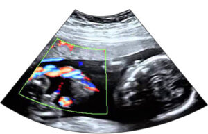
A fetal echocardiogram (fetal echo) is an ultrasound of your baby’s heart, typically done between the second and third trimester. It uses sound waves to create detailed images of the heart’s chambers, valves, and blood vessels. The test is painless and non-invasive, with no special preparation required. A gel is applied to your belly, and a sonographer uses a small device called a transducer to capture images of your baby’s heart.
The procedure usually takes about 60 minutes, though it may take longer if the baby’s position makes it harder to see. Fetal echocardiograms are often recommended if there’s a family history of heart problems, the mother has medical conditions like diabetes, or if a heart issue is suspected from other ultrasounds. This test helps identify congenital heart defects or heart rhythm problems.
The fetal echo allows early detection of heart issues, which can lead to lifesaving care after birth. While some small heart abnormalities might not be detected due to the baby’s developing heart, most minor issues do not cause problems at birth. The procedure is safe for both you and your baby. After the test, you’ll have a consultation to discuss the results, and a report will be sent to your doctor. This test provides valuable information to ensure the best care for your baby.
Doppler Ultrasound
Doppler ultrasound is used to look at the blood flow of your baby to check on his or her health. Different types of doppler tests are used based on medical indications and status of the pregnancy.